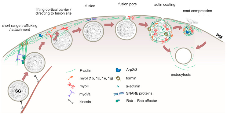
Figure 1.
A simplified model of the roles of actin and myosins in non-neuronal exocytosis.The scheme depicts potentially conserved or common mechanisms that have been observed in at least two different secretory cell types. Secretory vesicles are usually transported to the cell periphery on microtubules by motor proteins kinesins. Once in close proximity to the cell cortex, secretory vesicles might be guided towards (and attached to) the cortex by myoV motors along actin bundles attached to the PM. Actin cortex remodeling by myoII and possibly also myoI is necessary to enable access of large secretory vesicles to the PM for fusion of vesicles with the PM. The cortical actin network is essential to direct exocytic vesicles to the fusion site and for stable attachment or docking of granules close to the PM to facilitate fusion via SNARE proteins. F-Actin and myosin also provide force to modulate fusion pore diameters. Actin is polymerized on fused vesicles. Myosins are then recruited to the “actin coat” to either generate compressive forces for active secretion of vesicle contents or integration of vesicle membrane against a high hydrostatic pressure into the PM, likely in conjunction with an actin polymerization/crosslinking mechanism. In some cells, actin coats also stabilize fused secretory vesicles for endocytic retrieval.

