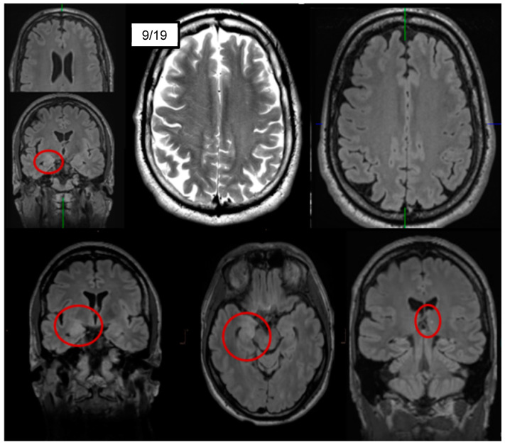
Figure 2.
Magnetic resonance imaging showing fluid-attenuated inversion recovery (FLAIR) hyperintensities right-mesio-temporally and on the right side of the corpus amygdaloideum, as well as a small, possibly microangiopathic, lesion in the left thalamus (bottom row). In addition, a slight grey–white matter blurring was observed (top center and right; cf. [8]).

