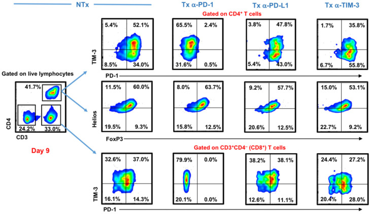
Figure 1.
Effect of different immune checkpoint inhibitors on T cells in breast tumor explants. Tumor tissue from 2 breast cancer patients were cut into small pieces and cultured with exogenous interleukin-2 (IL-2), in the presence or absence of anti-programmed cell death protein 1 (PD-1), anti-programmed death ligand-1 (PD-L1), or anti-T cell immunoglobulin and mucin-domain containing-3 (TIM-3) monoclonal antibodies (mAbs). Cells were collected on Day 9 and stained with TIM-3, PD-1, and different T regulatory cell (Treg)-related markers. Representative flow cytometric plots show TIM-3 and PD-1 surface expression on CD3+CD4− (CD8+) and CD3+CD4+ T cells, as well as intracellular FoxP3 and Helios expression on CD3+CD4+ T cells from different treatment conditions.

