Figure 2.
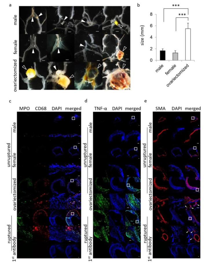
The exacerbation of the pathology of intracranial aneurysm in female rats with the bilateral ovariectomy. Ten-week-old female Sprague−Dawley rats were subjected to the aneurysm model. At 120 days after the surgical manipulations, specimens of induced lesions were harvested. (a,b) The macroscopic image of induced lesions (a) and their size (b) in each group; male rats (n = 18), female rats without ovariectomy (female, n = 10) or female rats with the bilateral ovariectomy (ovariectomized, n = 10). Bar, 1.0 mm (a). The white arrows and the black ones in (a) indicate the unruptured and the ruptured lesions, respectively. Bars in (b) indicate the mean ± SEM. Statistical analysis was done by the Tukey–Kramer method. ***; p < 0.001. (c,d) show similarity of unruptured lesions induced in female animals with the bilateral ovariectomy with ruptured ones. The representative images of immunostaining for a marker for neutrophil, myeloperoxidase (MPO, green in (c)), a marker for macrophage, CD68 (red in (c)), TNF-α (green in (d)), a marker for smooth muscle cell, smooth muscle alpha-actin (SMA, red in (e)), nuclear staining by DAPI (blue) or merged images are shown. The immunostaining without a 1st antibody served as a negative control and the representative images of this staining are shown in the lowest panels. Ruptured lesions were from female rats with the bilateral ovariectomy. The arrow in (e) indicates vasa vasorum with SMA-positive media. The magnified images, corresponding to a square in the upper panels, are shown in the lower panels. Bar, 20 μm.

