Figure 8.
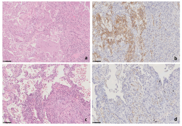
SPARC protein expression by immunohistochemical analysis. (a,b) Morphological features of case (LCCH-773) of lung adenocarcinoma, negative for SPARC promoter methylation, showing strong SPARC expression. (a) H&E staining, original magnification 20×; (b) SPARC staining, 20× original magnification. (c,d) Morphological features of case LCCH-889 of lung adenocarcinoma that does not show SPARC expression in tumor cells. (c) H&E staining, original magnification 20×; (d) SPARC staining, 20× original magnification. Small peritumoral vessels represent positive internal control. H&E—Hematoxylin and eosin stain.
SPARC protein expression by immunohistochemical analysis. (a,b) Morphological features of case (LCCH-773) of lung adenocarcinoma, negative for SPARC promoter methylation, showing strong SPARC expression. (a) H&E staining, original magnification 20×; (b) SPARC staining, 20× original magnification. (c,d) Morphological features of case LCCH-889 of lung adenocarcinoma that does not show SPARC expression in tumor cells. (c) H&E staining, original magnification 20×; (d) SPARC staining, 20× original magnification. Small peritumoral vessels represent positive internal control. H&E—Hematoxylin and eosin stain.

