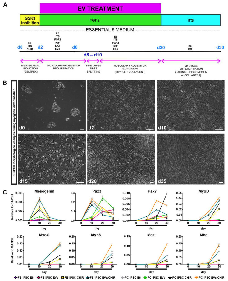
Figure 4.
Myogenic differentiation by MT-derived EVs. (A) Schematic diagram of the myogenic differentiation procedure: 30% confluent iPSCs were differentiated to early mesoderm using CHIR for 48 h, subsequently proliferation and expansion of muscular progenitors were stimulated utilizing EVs, FGF2, IGF, and the myotube-like cell maturation was induced, removing growth factors from the culture medium. (B) Morphological changes of PC-derived iPSC colonies during skeletal muscle differentiation. Scale bars represent 100 μm. (C) Representative qRT-PCR for the expression of early (Mesogenin, Pax3, Pax7, MyoD, MyoG) and late (MyH8, MCK, MHC) skeletal muscle genes. The analysis was performed at different time points (d0, d10, d20, d30) in the following conditions: FB- and PC-derived iPSCs not treated with EVs or CHIR (FB-iPSCs E6 and PC-iPSCs E6), exposed to CHIR or EVs only (FB-iPSC EVs, FB-iPSC CHIR, PC-iPSC EVs, PC-iPSC CHIR) and treated with both, EVs and CHIR (FB-iPSC EVs/CHIR and PC-iPSC EVs/CHIR) (n = 3 donors). Results were normalized to GAPDH.

