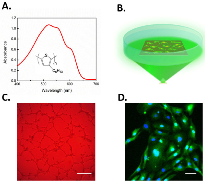
Figure 5.
(A) The rr-P3HT chemical structure and optical absorption of the thin film. (B) Experimental setup and optical excitation protocol performed for evaluation of polymer-mediated cell photoexcitation effects on cell fate. (C) Representative image of in vitro network formation in ECFCs seeded on P3HT and subjected to optical excitation; scale bar: 250 µM. (D) Representative image of immunofluorescence staining, showing enhanced light-induced NF-κB nuclear translocation. Cell nuclei are detected by DAPI (blue) while cytoplasmic p65 NF-κB subunit with a secondary chicken anti-rabbit Alexa(488)-conjugated antibody (green). Scale bar: 50 µM. Figures adapted from [44,207].

