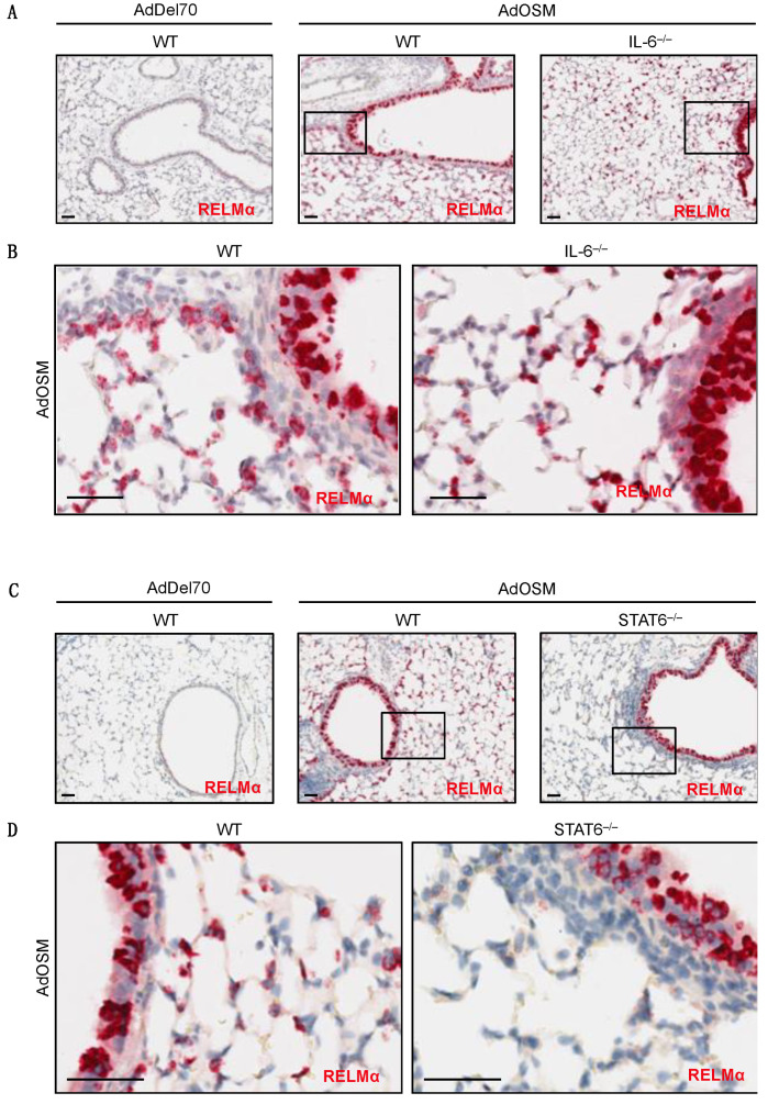
Figure 4.
RELMα mRNA induction in situ by OSM in IL-6–/– and STAT6–/– mice. Representative images are shown of CISH staining for RELMα (red) in formalin-fixed paraffin-embedded lung tissue sections from C57Bl/6 mice 7 days after treatment (n = 4–8 mice/group). (A) RELMα mRNA (red) in AdDel70-treated wild-type, AdOSM-treated wild-type or IL-6–/– mouse lung sections as indicated. (B) High magnifications images of boxed regions in A from AdOSM-treated mouse lungs of wild-type (left panel) or IL-6–/– (right panel). (C) RELMα mRNA (red) in AdDel70-treated wild-type, AdOSM-treated wild-type and STAT6–/– mouse lung sections as indicated. (D) Higher magnification images of indicated (boxed) regions from AdOSM-treated mouse lungs in (C). Scale bars, 50 µm.

