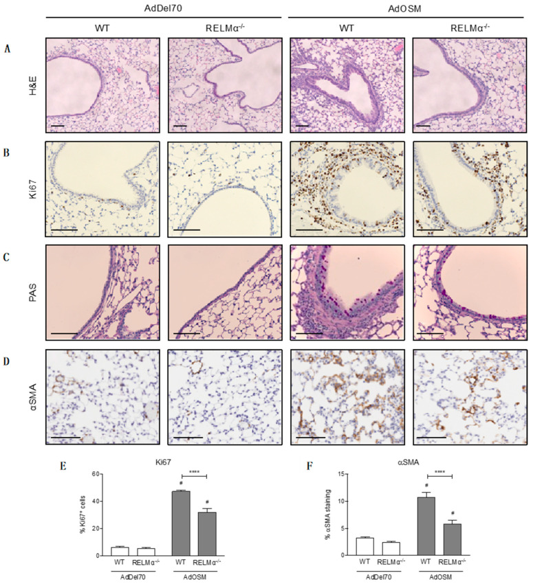
Figure 9.
Histological analyses of formalin-fixed, paraffin-embedded lung tissue. Lung sections were prepared from AdDel70- or AdOSM-treated wild-type or RELMα–/– mice as indicated. Representative images of lung tissue sections stained with (A) H&E, (B) Ki67 for proliferating cells, (C) PAS for mucous-producing goblet cells, and (D) αSMA. Scale bars, 100 µm. (E) Quantification of % Ki67+ cells in the epithelium and parenchyma. (F) Quantification of % αSMA staining in the parenchyma per lung tissue section, 3 sections were analyzed per mouse, excluding major airways and blood vessels. Data (E,F) are expressed as mean ± SEM, # different from AdDel70 (p < 0.05). Statistical significance was determined by two-way ANOVA with Tukey’s post hoc test (**** p < 0.0001 between indicated groups).

