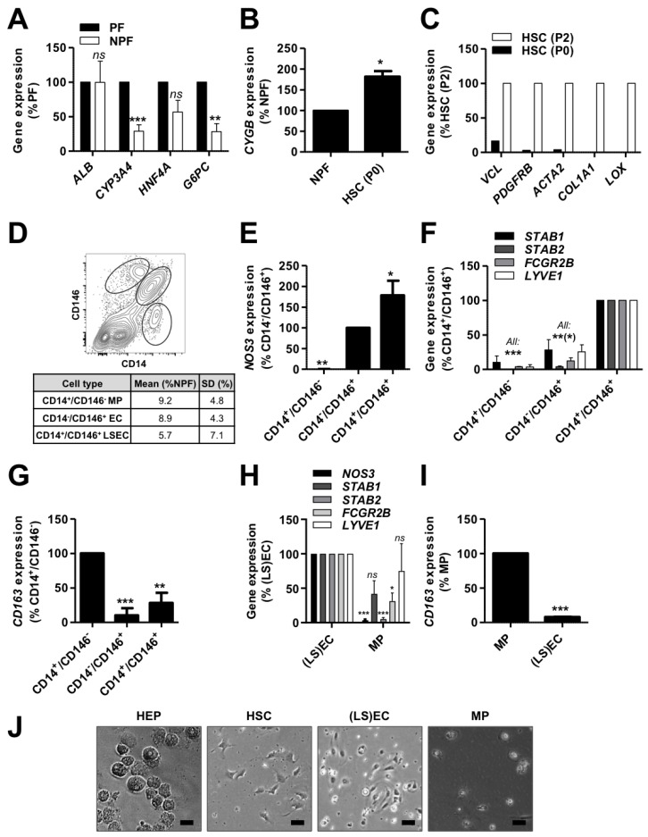
Figure 1.
Isolation of human primary liver cells. Human primary liver cells were isolated from cryopreserved material of 4 donors (155, 156, 164 and 173). Gene expression was evaluated by qPCR (A) in hepatocytes (HEPs, n = 4), (B) in hepatic stellate cells (HSCs) just after isolation (P0, n = 3) and (C) after 2 passages on tissue culture plastic (P2, n = 2) for representative markers. (D) Representative flow cytometry dot plot (upper panel) and average yields after non-parenchymal fraction (NPF) FACS according to CD146 and CD14 expression. (E–G) Gene expression was evaluated by qPCR in CD14+CD146− macrophages (MPs), CD14−CD146+ endothelial cells (ECs), CD14+CD146+ liver sinusoidal ECs (LSECs) and CD14−CD146− other cells for representative markers (n = 3). (H,I) After magnetic beads-activated cell sorting (MACS) for (i) CD146 and (ii) CD14, gene expression was assessed by qPCR in CD146+ (LS)ECs and CD146−CD14+ MPs for representative markers (n = 3−4). (J) Representative pictures of freshly isolated cells (scale bar = 50 μm). All: results were analyzed per donor and are expressed as percentage of expression in the indicated fraction (expected maximal expression), graphs show mean ± SEM, Student’s t-test (A,B and H,I) or one-way ANOVA with Dunnett’s post-hoc test (E–G), ns p > 0.05, * p < 0.05, ** p < 0.01, *** p < 0.005. ACTA2: actin α2, ALB: albumin, COL1A1: collagen type 1 α1 chain, CYGB: cytoglobin, CYP3A4: cytochrome P450 3A4, FCGR2B: Fcγ receptor 2b, G6PC: glucose 6 phosphatase catalytic subunit, HNF4A: hepatocyte nuclear factor 4 α, LOX: lysyl oxidase, LYVE1: lymphatic vessel endothelial hyaluronan receptor 1, NOS3: NO synthase 3, PDGFRB: PDGF receptor β, STAB: stabilin, VCL: vinculin.

