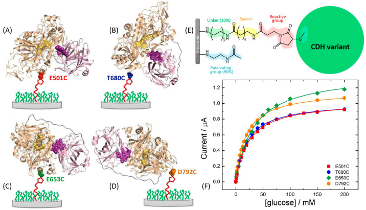
Figure 13.
Cartoon representations of the structures of the four different MtCDH variants attached to the electrode surface in different orientations through the cysteine-maleimide bond. The cytochrome domain is shown in purple and the dehydrogenase domain in pale brown. (A) E501C, substrate channel close to electrode (front on); (B) T680C, top of enzyme facing electrode (top on); (C) E653C, right side of substrate channel facing electrode (right side on); and (D) D792C, C-terminus close to electrode (bottom on). The images were obtained with PyMol software based on the crystal structure of MtCDH, PDB code 4QI6. (E) Chemical structure of the whole electrode modification used in this work to immobilize MtCDH variants with a single surface exposed cysteine, with the different components in different colors. (F) Background-corrected catalytic currents at 0.0 V vs. SCE for the four MtCDH variants from parts (A–D) plotted as a function of the glucose concentration. The definition of all used mutants and their abbreviate names can be found in ref. [167]. (The figure was adopted from ref. [167] with permission.)

