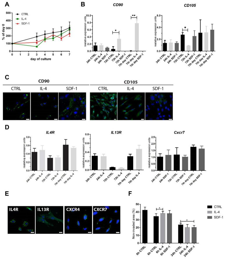
Figure 1.
Characterization of mouse adipose tissue-derived stromal cells (mADSCs) cultured under control conditions or in the presence of IL-4 or SDF-1. (A) Growth curves of mADSCs cultured for 7 days; data shown as a proportion of the number observed at day 0. (B) Analysis of the level of mRNAs encoding CD90 and CD105. Expression was related to the levels observed in control cells at day 0 (beginning of the culture) and normalized to mRNA encoding hypoxanthine phosphoribosyl transferase, i.e., HPRT. (C) Localization of CD90 or CD105 (green) and nuclei (blue) in mADSCs after 72 h of culture, bar = 20 µm. (D) Analysis of the level of mRNAs encoding IL4R, IL13R, and CXCR7. Expression was related to the levels observed in control cells at day 0 (beginning of the culture) and normalized to mRNA encoding HPRT. (E) Localization of IL4R, IL13R, CXCR7, or CXCR4 (green) and nuclei (blue) in control mADSCs after 7 days of culture, bar = 20 µm. (F) In vitro migration assay—mADSCs were scratched from the culture dish and the area which was not invaded by migrating cells was measured and presented as the proportion (%) of the whole area photographed (0 h, 6 h, and 24 h). For each experimental group n ≥ 3. Data are presented as mean ± SD. Data have been analyzed using Student’s t-test (A); two-way ANOVA test (d-CXCR7, F); Kruskal-Wallis test (B,D—IL4R, IL13R). * p < 0.05; ** p < 0.01.

