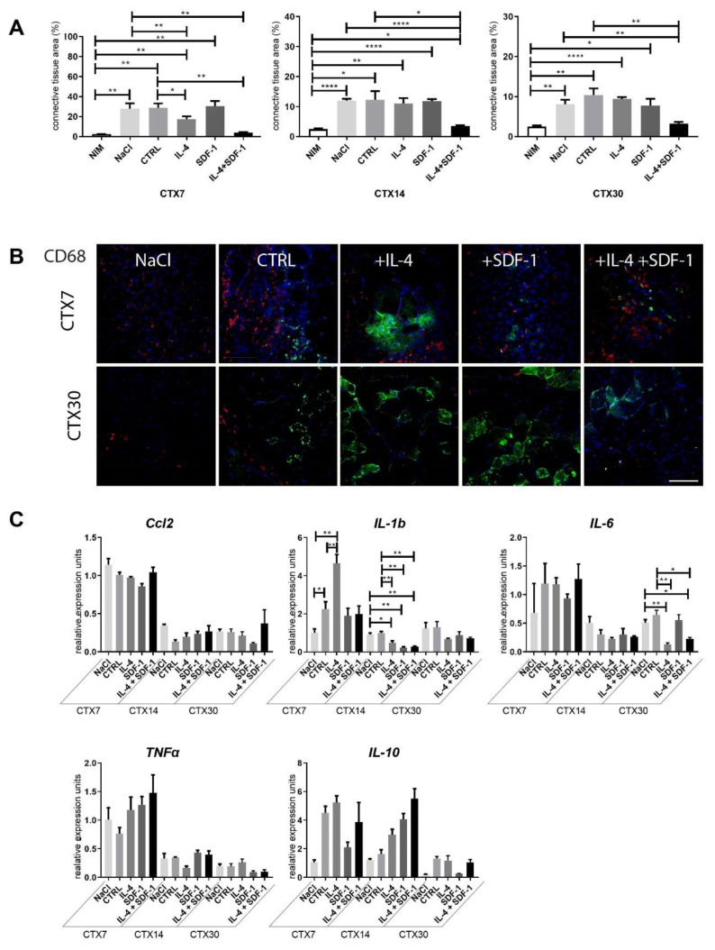
Figure 4.
Connective tissue formation and immune response of mouse skeletal muscles which received NaCl, control mADSCs, or mADSCs pretreated with IL-4, SDF-1, or IL-4 and SDF-1. (A) Connective tissue area calculated from the histological sections (Masson’s Trichrome staining) analyzed at day 7, 14, and 30 of regeneration. (B) Localization of CD68+ cells and ADSCs in regenerating skeletal muscles assessed at day 7 and 30 of regeneration. CD68 (red), GFP (green), nuclei (blue), bar = 100 µm. (C) Expression of mRNAs encoding Ccl2, IL-1b, IL-6, TNFα, and IL-10, analyzed at day 7, 14, and 30 of regeneration. Expression was related to the mean expression level in control muscle analyzed at day 7 and normalized to mRNA encoding HPRT. CTX7 - day 7; CTX14 - day 14; CTX30 - day 30 of regeneration, For each experimental group n ≥ 3. Data are presented as mean ± SD. Data have been analyzed using two-way ANOVA test (a, c—IL-1b, IL-6); Kruskal-Wallis test (c-Ccl2, TNFα, IL-10). * p < 0.05; ** p < 0.01, *** p < 0.001, **** p < 0.0001.

