Figure 4.
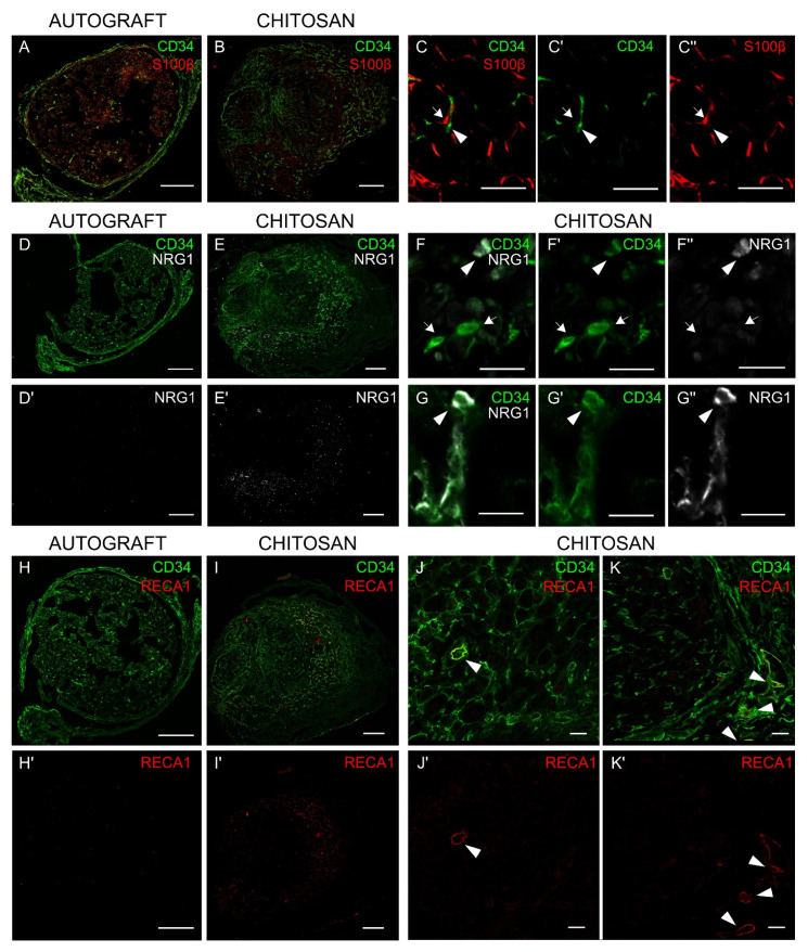
Nerve fibroblasts express NRG1 in vivo. Cross section of median nerves analyzed 7 days after repair with an autograft (A, D, D’, H, H’) or with a chitosan conduit (B, E, E’, I, I’): (A,B) double-stained with S100β (Schwann cell marker, red) and CD34 (fibroblast marker, green). (C, C’, C’’) Images at higher magnification show the absence of colocalization between S100β and CD34; fibroblasts are pointed out by arrow heads, Schwann cells by arrows. (D,E) double-stained with CD34 (green) and NRG1 (white) or (D’,E’) single-stained with NRG1; (D’) and (E’) highlight different levels of NRG1 expression in the two different experimental models (higher in the chitosan group). (F, F’, F” and G, G’, G”) Images at higher magnification show some CD34+ cells positive only to CD34 (arrow) and some CD34+ cells also positive to NRG1 (arrow head). (H,I) double-stained with CD34 (fibroblast and endothelial cell marker, green) and RECA1 (endothelial cell marker, red) or (H’,I’) single-stained with RECA1; (H’) and (I’) highlight different levels of RECA1 expression in the two different experimental models (higher in the chitosan group). (J, J’ and K, K’) Images at higher magnification show some CD34+ cells also positive for RECA1, corresponding to endothelial cells (arrow heads), and several CD34+ cells positive only to CD34, corresponding to fibroblast cells. Scale bars: 200 µm (A, B, D, D’, E, E’, H, H’, I, I’); 20 µm (J, J’, K, K’); 10 µm (C,C’,C’’, F, F’, F’’, G, G’, G’’).

