Figure 1.
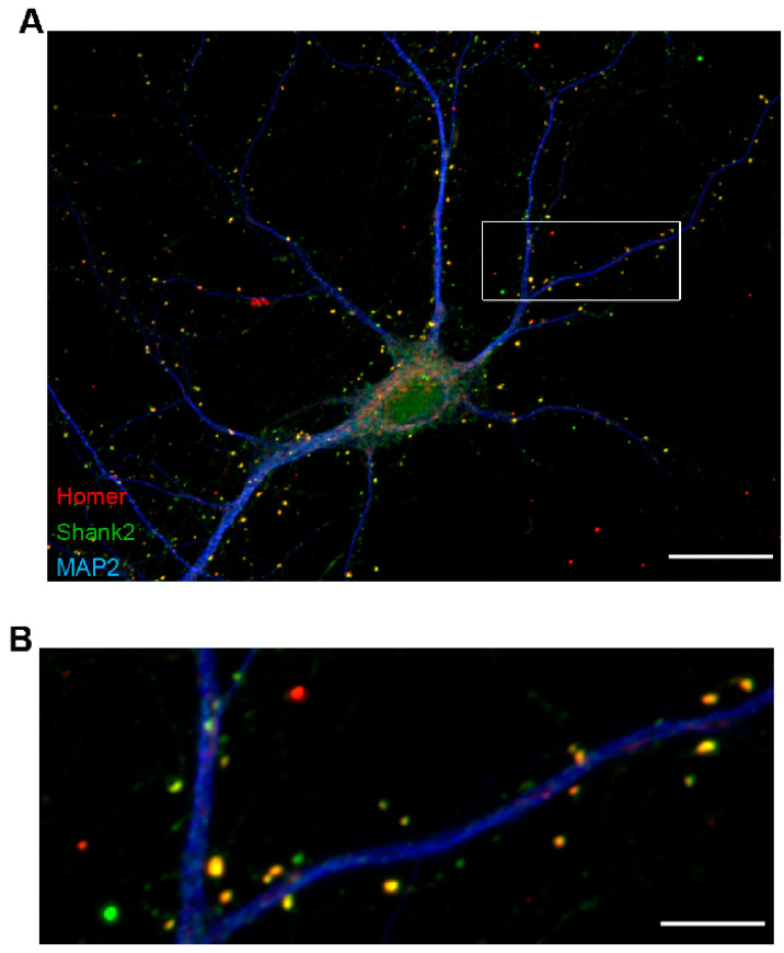
Single cell layer representation of postsynaptic proteins Homer and Shank2/ProSAP1. (A) Immunostaining of cultured rat hippocampal neurons (DIV16) recorded with a 63×/1.4 objective on a Zeiss AxioObserver.Z1 equipped with an ApoTome.2 confirms colocalization of the postsynaptic density proteins Homer 1 (Homer; red) and postsynaptic density protein Shank2/ProSAP1 (Shank2; green) at structures protruding from dendrites marked by microtubule-associated protein 2 (MAP2; blue). (B) Enlarged view of the boxed region depicted in A. Scale bar, 20 µm in (A), 5 µm in (B).

