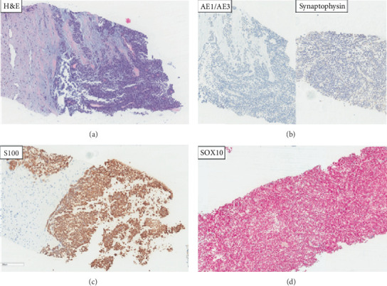
Figure 2.

H&E (a) shows loosely discohesive epithelioid cells with eosinophilic to clear cytoplasm, round to ovoid nuclei, and prominent nucleoli with a solid growth pattern. Keratin AE1/AE3 and synaptophysin IHC negative (b). S100 (c) and SOX10 (d) IHC diffusely positive. Red chromogen IHC showing positive red staining in the nuclei.
