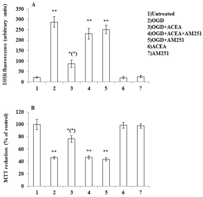
Figure 4.
CB1 stimulation with arachidonyl-2′-chloroethylamide (ACEA) prevents OGD-induced ROS formation as well as toxicity in primary cortical neurons. (A) DHR-loaded cells, were incubated for 15 min with AM251 (0.5 µM), exposed to ACEA (0.5 µM) for an additional 15 min and, finally, subjected to OGD (60 min). After 1.5 h incubation under normoxic conditions, the cells were analyzed with a fluorescence microscope as described in Figure 1. Results expressed as arbitrary units represent the mean ± SEM calculated from three to five separate experiments, each performed in duplicate. * p < 0.05, ** p < 0.001 vs. control cells; (*) p < 0.001 vs. OGD-treated cells (one-way ANOVA followed by Bonferroni test). (B) Neurons were treated as in (A), exposed to OGD for 60 min, and after 20 h under normoxic conditions, were analyzed for cell viability using the MTT assay. Results represent the mean ± SEM of three to five separate experiments, each performed in duplicate. * p < 0.05, ** p < 0.001 vs. control cells; (*) p < 0.01 compared to OGD-treated cells (one-way ANOVA followed by Bonferroni test).

