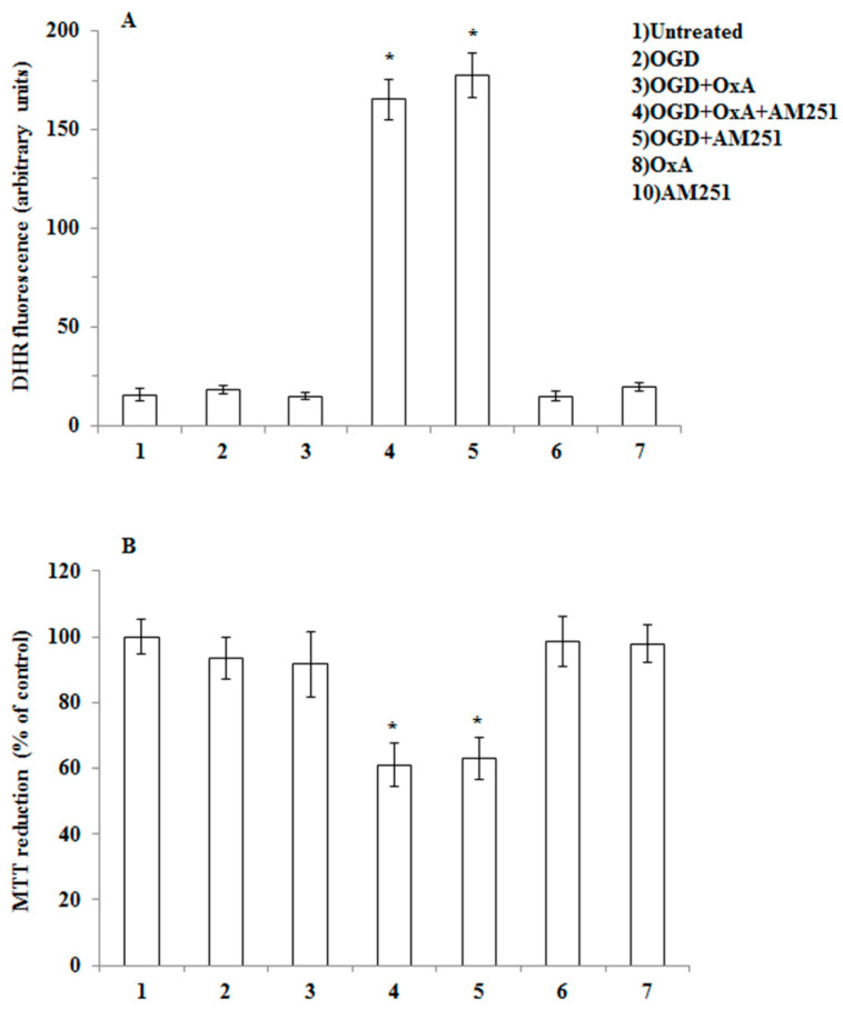
Figure 5.
OGD treatment fails to induce ROS formation and toxicity in primary cortical neurons isolated from MAGL null mice. (A) Primary neurons isolated from MAGL null mice, loaded with DHR, were incubated with AM251 (0.5 µM, 15 min), exposed to OX-A (0.2 µM) for an additional 30 min and, finally, subjected to OGD for 60 min. After 1.5 h incubation under normoxic conditions, the cells were analyzed with a fluorescence microscope as described in Figure 1. Results expressed as arbitrary units represent the mean ± SEM calculated from three to five separate experiments, each performed in duplicate. * p < 0.0001 vs. OGD-treated cells (one-way ANOVA followed by Bonferroni test). (B) Neurons were treated as in (A), exposed to OGD for 60, and after 20 h under normoxic conditions, analyzed for cell viability using the MTT assay. Results represent the mean ± SEM of three separate experiments, each performed in duplicate. * p < 0.01 vs. OGD-treated cells (one-way ANOVA followed by Bonferroni test).

