Figure 3.
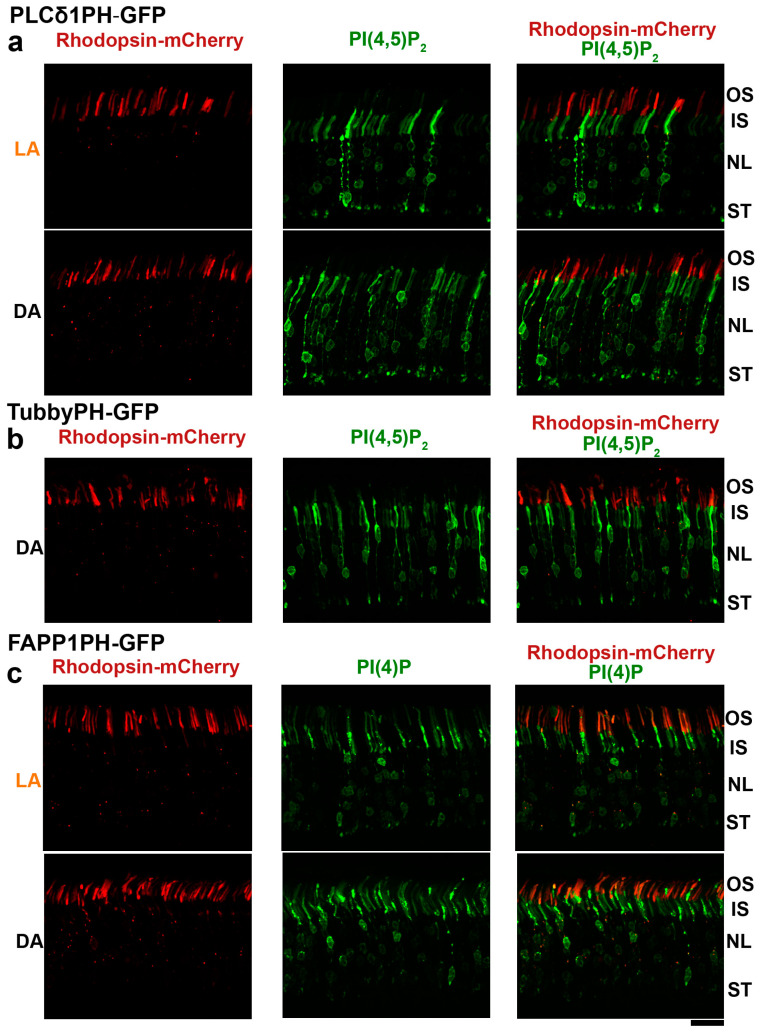
Distribution of PI(4)P and PI(4,5)P2 phosphoinositide sensors in rod photoreceptors. Green Fluorescent Protein (GFP) fusion constructs of PI(4,5)P2-binding domains from PLC δ1 (a) and Tubby (b) proteins and the PI(4)P-binding domain of FAPP1 (c) were electroporated at P0 into rod photoreceptors to investigate the localization of corresponding phosphoinositides. Their distribution patterns (green) were analyzed in retinal cross-sections of 6-week-old dark (DA)- or light (LA)- adapted mice. Rhodopsin fused with mCherry (red) was used as a rod outer segment marker. PI(4,5)P2 sensors produce intense signals in the inner segments (IS), nuclear layer (NL) and synaptic terminals (ST), but are virtually excluded from outer segments (OS). The PI(4)P sensor shows signal in the outer segments, around nuclei, and in the inner segments. Occasional red puncta detected in the inner segments most likely represent mistargeted Rhodopsin-mCherry fusion protein. Scale bar: 20 μm.

