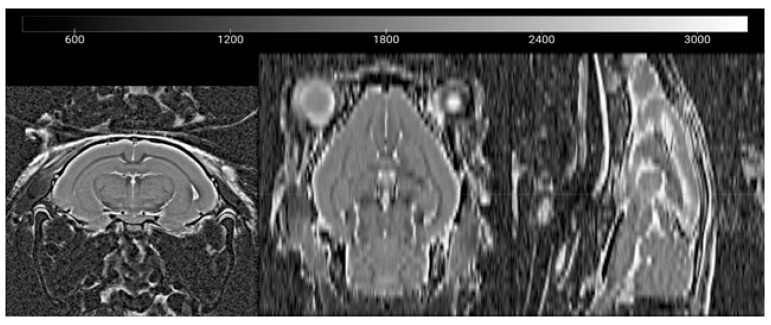
Figure A1.
Example of T2 Volumetric Images. Representative T2-weighted image showing coronal, sagittal, and axial views of an adolescent guinea pig brain. Left–right orientation is as shown in the coronal image, the anterior (rostral) portions of the brain face upwards in the sagittal and axial views. Image created in MRIcroGL v1.2.

