Figure 6.
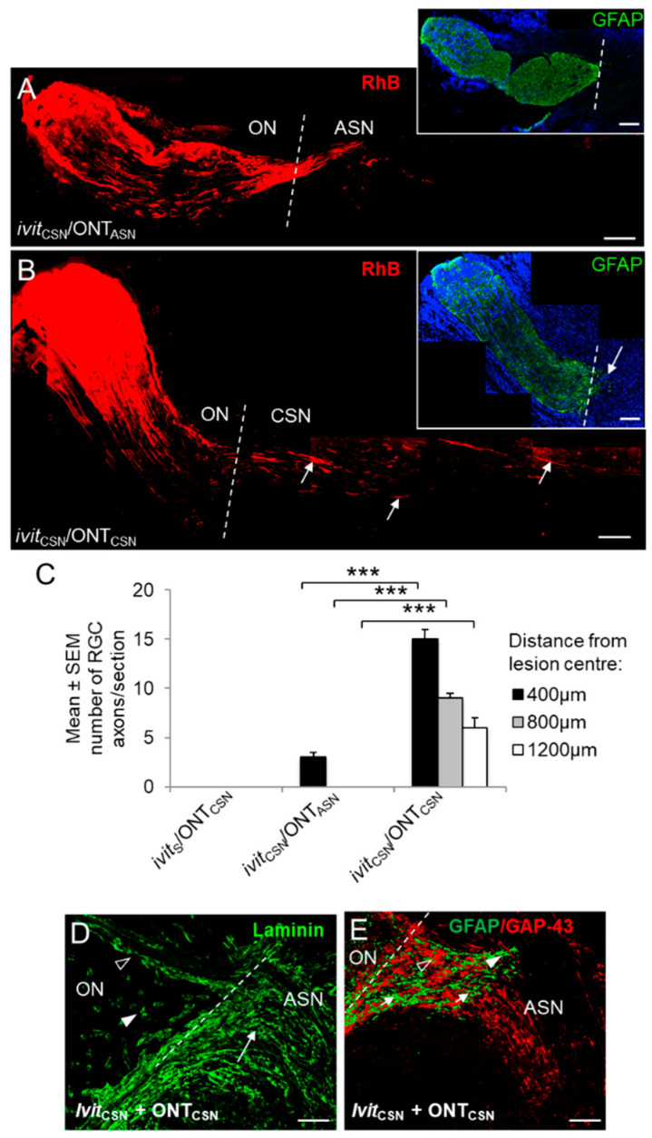
Regeneration of rhodamine B (RhB)+ RGC axons (red) into cellular sciatic nerve (CSN) or acellular sciatic nerve (ASN) grafts at 21 days after optic nerve transection (ONT). Representative images of longitudinal sections of optic nerve and graft in (A), ivitCSN/ONTASN and (B), ivitCSN/ONTCSN groups—note the presence throughout the length of the ONTCSN graft of RhB+ (red) regenerating RGC axons (open arrows) and their limited penetration into the ONTASN graft (A,B insets), optic nerve GFAP+ astrocyte processes (green) do not enter ONTASN grafts, but do enter the proximal region of the ONTCSN graft (closed arrow) (scale bars in A, B, and D = 200 µm; broken line indicates anastomosis site). (C) Quantification of the number of axons extending 400, 800, and 1200 μm past the anastomosis site into the ONTCSN graft (*** p < 0.0001). A few axons penetrate the junctional zone of ONTASN grafts compared to the numbers extending for long distances into ONTCSN grafts. (D), anastomosis site in an ivitCSN/ONTCSN rat showing the laminin+ basal lamina of the optic nerve vasculature (arrow heads) and Schwann cell basal lamina tubes aligned in parallel arrays in the ONTCSN graft (arrow), with which (E) regenerating RGC axons are associated with the extent of GFAP+ astrocyte process (green) invasion and the growth of RhB+ RGC axons (red) (scale bars = 150 µm; the broken line indicates the anastomosis site).

