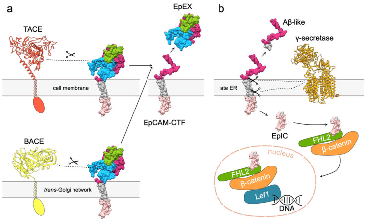
Figure 5.
EpCAM signaling via RIP. (a) Cleavage of EpCAM extracellular part by either TACE or BACE results in release of soluble EpEX and membrane bound EpCAM-CTF. Extracellular part of TACE was modeled using Robetta [53,54], and structure extracellular part of BACE was obtained from the Protein Data Bank (PDB 2WJO). Transmembrane and cytosolic regions of both proteases are depicted schematically. EpCAM is presented as molecular surface; transmembrane region and intracellular domain are shown in gray and light pink, respectively. (b) Cleavage of EpCAM-CTF by γ -secretase complex (PDB 5A63) results in release of Aβ-like peptide and EpIC that is recruited in the EpIC–FHL2–β-catenin–Lef1 signaling complex. FHL (green), β-catenin (orange) and Lef1 (blue) are depicted by shapes corresponding to their relative sizes.

