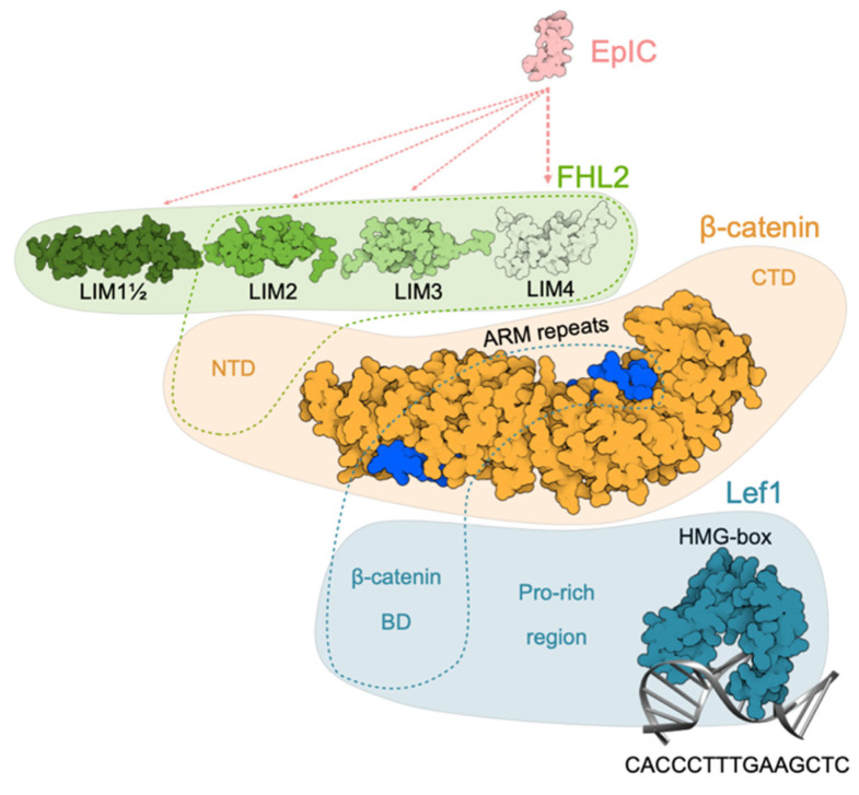
Figure 7.
Schematic model of EpIC–FHL2–β-catenin–Lef1 signaling complex. EpIC was modeled using MODELLER [64]. Binding of EpIC to FHL2 is indicated by dotted lines (light pink; width is related to importance of interaction). First and a half, second, third and fourth domain of FHL2 are depicted based on corresponding NMR structures (PDB 2MIU, 1X4K, 2D8Z, and 1X4L respectively). Binding of FHL2 to β-catenin N-terminal domain is indicated by a green dotted outline. β-catenin is represented by structure of ARM repeats with bound part of Lef1 β-catenin BD (PDB 3OUW) and relative positions of N- and C-terminal domains (NTD and CTD, respectively), the structures of which are yet unknown. Position of β-catenin BD is indicated by blue dotted outline. Structure of Lef1, except for the C-terminal HMG-BOX bound to its target DNA sequence (PDB 2LEF), is not known. β-catenin BD and Pro-rich region are indicated at their relative position.

