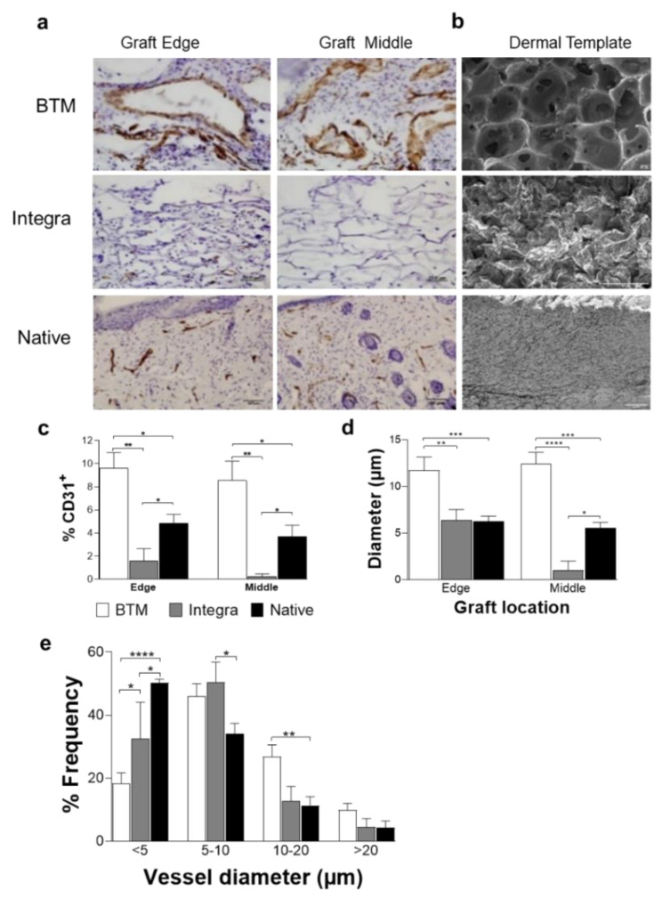
Figure 1.
Detection of endothelial cells in mouse grafts. (a) CD31 positive endothelial cells were detected in grafts using IHC staining. Representative images of BTM, Integra®, and allogenic native skin grafts are presented (scale bar 50 µm). (b) SEM images of BTM, Integra®, and native mouse skin (scale bar 100 µm). (c) CD31 staining on graft edge and the middle was quantified and normalised for image area. CD31 staining was used to score vessel diameter (d), and the frequency of different vessel diameters (e). Values represent mean +/- SEM in each group (n = 4 mice per group) and analysed using unpaired t-test. * = p ≤ 0.05, ** = p ≤ 0.01, *** = p ≤ 0.001, **** = p ≤ 0.0001.

