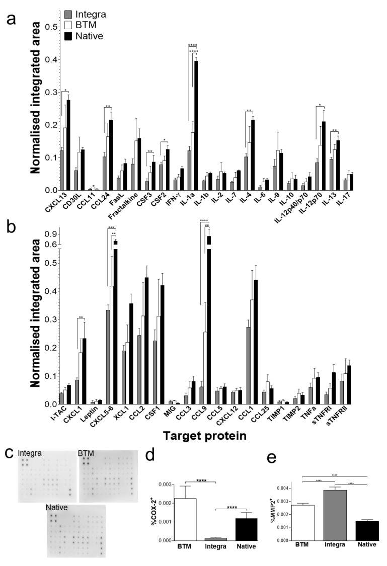
Figure 4.
Protein expression profiling of 2-week-long grafts. (a,b) Protein signals recorded for each target using 10-second exposure and normalised to positive controls. Mean and SEM values presented for each target (n = 4 per group) (a) Targets from rows 1 and 2 position F6 to rows 5 and 6 position B2 of the array). (b) Targets from rows 5 and 6 position B3 to rows 7 and 8 position I9 of the array. The full map of the array is provided in Supplementary Data Table S1. (c) Representative chemiluminescent protein array blots for each group. (d) Quantification of immunoperoxidase staining for inflammation marker, COX-2 (n = 4–8 mice per group), using unpaired t-test * = p < 0.05, ** = p < 0.01, *** = p < 0.001, **** = p < 0.0001. (e) Quantification of immunoperoxidase staining for ECM remodelling enzyme, MMP-2 (n = 4–8 mice per group). Results analysed using unpaired t-test, significant p-values as for panel c. Abbreviations: CCL1 (TCA-3), chemokine CC motif ligand 1; CCL2 (MCP-1), chemokine CC motif ligand 2; CCL3 (MIP-1α), chemokine CC motif ligand 3; CCL5 (RANTES), chemokine CC motif ligand 5; CCL9 (MIP1γ), chemokine CC motif ligand 9; CCL11 (Eotaxin), chemokine CC motif ligand 11; CCL24 (Eotaxin-2), chemokine CC motif ligand 24; CCL25 (TECK), chemokine CC motif ligand 25; CSF1 (M-CSF), colony-stimulating factor 1; CSF2 (GM-CSF), colony stimulating factor 2; CSF3 (G-CSF), colony stimulating factor 3; CXCL1 (KC), chemokine CXC motif ligand 1; CXCL5-6 (LIX), chemokine CXC motif ligand 5-6; CXCL12 (SDF-1), chemokine CXC motif ligand 12; CXCL13 (BLC), chemokine CXC motif ligand 13; IL1 or 6 or 10, interleukin 1 or 6 or 10; TIMP, tissue inhibitors of metalloproteinases; TNF, tumour necrosis factor, XCL1, lymphotactin α.

