Figure 5.
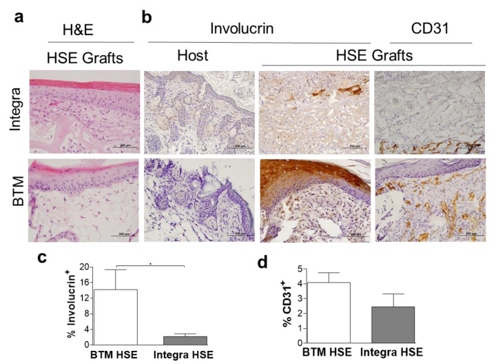
Neo-epidermis detection in 2-week-long HSE grafts. (a) Representative H&E staining of HSE constructed using Integra® (single layer) or BTM (single layer) two weeks post grafting (scale bar 100 µm). (b) Representative images of human-specific involucrin staining of mouse host and HSE grafts and CD31 in vivo (scale bar 100 µm). (c) Human involucrin staining of HSE grafts was quantified and Mean and SEM values are presented (n = 5–6 per group, *p value < 0.05). (d) CD31 expression was also quantified in HSE grafts. Mean and SEM values are presented (n = 5–6 per group).

