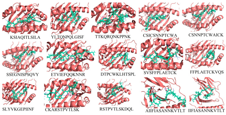
Figure 2.
The 3D binding pattern of the selected 15 epitopes (deep-salmon cartoon representation) docked with their respective HLAs (green-cyan sticks representation) as shown in Table 2. Hydrogen bond interactions are highlighted with blue color dotted lines.

