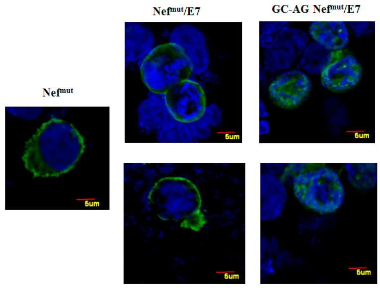
Figure 2.
Intracellular localization of both Nefmut/E7 and GC-AG Nefmut/E7. Confocal microscope analysis of HEK-293T cells transfected with either Nefmut, Nefmut/E7, or GC-AG Nefmut/E7 expressing vectors. Shown are representative fields from transfected cell cultures incubated first with an anti-Nef mAb, and then with Alexa 488-conjugated anti-mouse IgGs. DAPI (blue fluorescence) was used to highlight cell nuclei. Scale bars are reported. The results are representative of two independent experiments.

