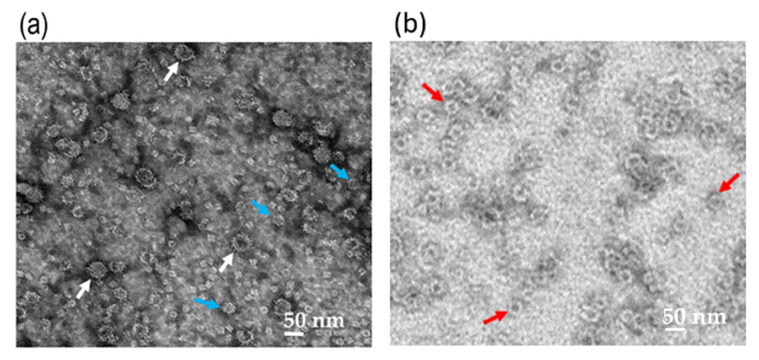
Figure 2.
Transmission electron micrograph of the Nc-aD protein. Transmission electron micrographs of the Sf9-produced Nc-aD protein (a), and E. coli-produced Nc-aD protein (b). Transmission electron microscopic analysis showed that the Sf9-produced Nc-aD protein assembled into virus-like particles (VLPs) ranging from ~21 nm (blue arrows) to ~55 nm (white arrows) in diameter, while the E. coli-produced Nc-aD protein assembled into VLPs of ~30 nm (red arrows) in diameter. The white bars indicate 50 nm.

