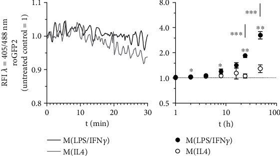
Figure 1.

Redox status of macrophages during M(LPS/IFNγ) vs. M(IL4) polarization. J774A.1 cells were transduced by vectors encoding the redox-sensitive marker proteins roGFP2. To mimic macrophage polarization, cells expressing roGFP2 were treated with 1 μg/ml LPS combined with 10 U/ml IFNγ (M(LPS/IFNγ)) or with 10 ng/ml IL4 (M(IL4)). The redox status was determined by FACS analysis at 405 nm and 488 nm during the first 30 min ongoing (left panel, representative experiment) or at indicated hours (right panel, quantitative data) after polarization starts. All experiments were performed at least three times. Mean values ± SD are provided. Untreated control cells were set as 1. (∗p ≤ 0.05, ∗∗p ≤ 0.01, ∗∗∗p ≤ 0.001).
