Figure 6.

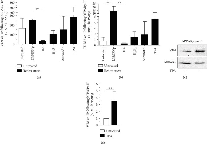
Protein-protein interaction of redox-modified hPPARγ. The LC/MS data sets of HA-immunoprecipitated HA-tagged hPPARγ with and without redox stress treated J774A.1 cells (1 μg/ml LPS combined with 10 U/ml IFNγ for 4 h, 10 ng/ml IL4 for 4 h, 100 μM H2O2 for 15 min, 1 μM TPA for 30 min) were reanalysed for hPPARγ coimmunoprecipitated proteins. Quantification of identified VIM (a) and TUBB5 (b) content via LC/MS is depicted. To verify LC/MS data, J774A.1 cells producing HA-tagged hPPARγ were treated with TPA (1 μM, 30 min) and coimmunoprecipitation for HA-hPPARγ was performed. Lysates of untreated cells were processed in parallel. All experiments were performed at least three times. (c) shows a representative Western blot analysis and quantification of mean values ± SD is provided in (d). (∗∗p ≤ 0.01).
