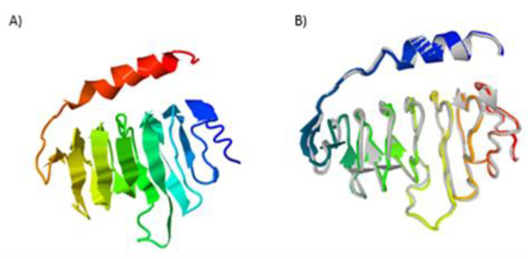
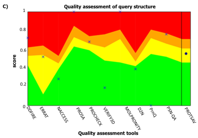
Figure 3.
Protein modelling, refinement and validation. (A) The final 3D model of OvMANE1 chimeric antigen gotten after homology modelling on I-TASSER. (B) Refined 3D structure overlay (colored) on the ‘crude model’ (gray) by GalaxyRefine. (C) Refined model validation using ProTSAV predicted the refined structure to be within the range of 2–5 Å estimated root mean squared deviation (RMSD).


