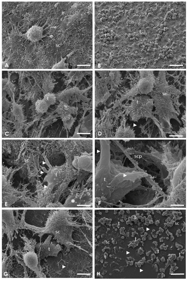
Figure 2.
Scanning electron micrographs of the interaction of A. culbertsoni trophozoites with Schwann cells (SC) at different point times (1, 2, 3, and 4 h). (A) No morphological evidence of damage was observed in the control culture. Bar = 10 μm. (B) After 1 h of coincubation, trophozoites (t) were observed to be adhered to the SC surface without cell damage. Bar = 10 μm. (C–E) After 2 h of co–incubation. (C) Trophozoite (t) in suggestive cell division. Bar = 10 μm (D) Trophozoites (t) were observed migrating beneath the SC cultures, evidencing the substrate (arrowhead). Bars = 10 μm. (E) Trophozoite (t) in contact with SC emitting sucker–like structures (arrowheads). A trophozoite migrating (asterisk) with its typical acanthopods was also observed. Bar = 1 μm. (F–G) After 3 h of co–incubation. (F) Amoebae were not only in contact with the cell body but were also in contact with the cytoplasmic prolongations of SC (scp) by sucker-like structures (arrowhead). Bar = 1 μm. (G) Trophozoites (t) in areas close to the substrate. Culture damage was evident (arrowhead). Bar = 1 μm. (H) After 4 h of co-incubation. Extensive lytic zones (arrowhead) were observed in SC cultures by Acanthamoeba trophozoites (t). Bar = 10 μm.

