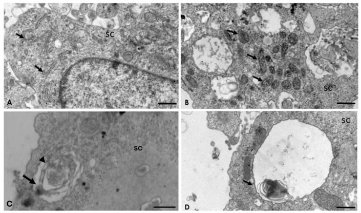
Figure 5.
Interaction of A. culbertsoni and Schwann cells (SC) after 2 h of interaction. (A,B) Ultrastructural alterations in organelles of SC were frequently observed (arrows) in the rough endoplasmic reticulum (A) and mitochondria (B) Bars = 2 µm. (C,D) Multilamellar bodies with cytoplasmic material (arrows) persisted. In electron micrograph (C) Bar = 500 nm, a double membrane (arrow head) is evident, characteristic of these structures. Bar = 2 µm.

