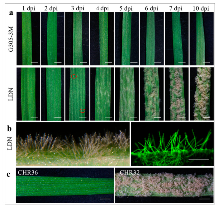
Figure 1.
Macroscopic and microscopic observations of the resistant and susceptible host plants at different time points after Bgt#70 inoculation. (a) Macroscopic observations of G305-3M and LDN at eight time points after Bgt#70 inoculation. The first colonies of Bgt are marked with a red circle at 3 dpi on LDN; (b) colony observation on LDN at 5 dpi after Bgt#70 infection. The Bgt colony on the left side was observed under light microscopy, while the colony on the right was observed under fluorescence microscopy after staining with wheat germ agglutinin (WGA); (c) macroscopic observations of the resistant RIL CHR36 and susceptible RIL CHR32 at 10 dpi. Scale bars: (a,c) 2 mm; (b) 250 μm.

