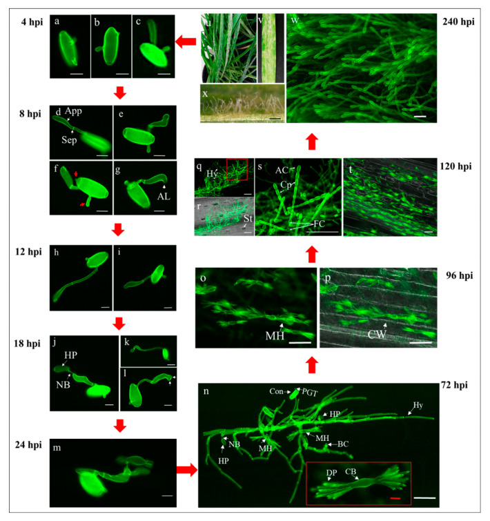
Figure 2.
Disease cycle of Bgt#70 on leaf segments of LDN (susceptible parent). All samples were stained with WGA prior to observation under fluorescence microscope; (a–c) germination of Bgt conidia at 4 hpi; (d–g) initial stages of appressorial germ tube formation at 8 hpi; (h,i) advanced stages of appressorial germ tubes formation at 12 hpi; (j–l) initial stages of penetration and invasion of Bgt into wheat cells at 18 hpi; (m) development of mature haustoria at 24 hpi; (n) formation of Bgt colonies at 72 dpi. A high-resolution close-up image of the mature feeding structure of Bgt with central body and digitate processes (finger-like projections) is shown in the red box; (o,p) feeding structures of Bgt at 96 hpi (4 dpi). Bgt haustorium within the invaded plant host cell under fluorescence microscopy (o) and superimposition of fluorescence and bright field images with visible multiple haustoria with multi-digits in individual epidermal cells (p); (q–t) images of massive colonization and reproduction of Bgt on wheat leaves at 120 hpi (5 dpi) observed under fluorescence microscopy (q,s,t) and superimposed on a bright field images; (r) colonies with numerous conidiophores and haustoria are shown. The high-resolution close-up image is derived from the red box in q; (u–x) observation of Bgt symptoms and conidiophores developed at 240 hpi (10 dpi). Powdery mildew symptoms on intact leaves (u) and leaf segments (v), observation under fluorescence microscopy of Bgt conidiophores, aerial hyphae under light microscopy. AL: apical lobe; App: appressorium; AC: apical conidium; CB: central body; Con: conidium; Cp: conidiophores; CW: cell wall; DP: digitate processes (finger-like projections); BC: bulbous conidiophore; MH: mature haustorium; HP: haustorial primordium; Hy: hyphae; NB: neckband; Sep: septum; St: stomata. Scale bars: (a–m) and (n) red, 10 µm; (o–t) and (n) white, 50 µm; (w), 100 µm; (x), 250 µm.

