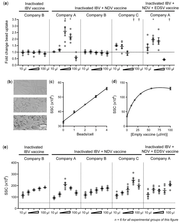
Figure 4.
Phagocytosis capacity of HD11 cells can be increased upon exposure to inactivated viral w/o vaccines. (a) The fold change in bead uptake by HD11 cells upon stimulation with vaccines is compared to unstimulated controls. The x-axis shows the graded doses at which the vaccines have been added, expressed as μL dose, added to 1 mL of cell culture medium. † indicates that datapoints were missing because the threshold of ≥100 viable cells was not reached. (b) Light microscopy photos show unstimulated HD11 cells (top), HD11 cells exposed to 10 μL/mL inactivated bivalent vaccine B (middle), and 100 μL/mL inactivated bivalent vaccine B (top). (c) A linear correlation curve shows the relationship between the average flow cytometric SSC and number of IgY-opsonized beads/cell for HD11 cells containing 0–4 beads/cell. (d) A non-linear saturation curve shows the relationship between the average SSC and different doses of empty vaccine (without viral antigens) from company B. (e) The flow cytometric SSC of HD11 cells is shown for graded doses of the different vaccines. Three independent experiments were performed, and the experimental conditions of each independent experiment were tested in duplicate. Error bars represent the SEM. The experimental groups were tested for statistically significant differences in bead uptake and SSC between stimulated and unstimulated groups using Kruskal–Wallis tests and Dunn’s multiple comparisons tests. Statistical significance is indicated by * p < 0.05. For figure (e), all data was found to be statistically different from the unstimulated sample with p < 0.001.

