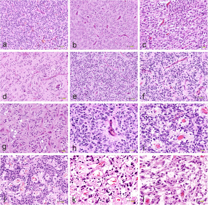
Fig. 7.
Representative histopathology of CNS-HGNT BCOR tumors. a Solid, highly cellular, monomorphic growth pattern with rich, thin-walled capillary network. b Pleomorphic cells of astroglial-like appearance. c Oligodendroglial-like monomorphic cells with round nuclei surrounded by clear cytoplasm accompanied by chicken-wire pattern of vessels. d Infiltration of uniform neoplastic cells with round nuclei and microcalcifications. e Densely-packed, small, hyperchromatic cells and thin-walled capillary blood vessels. f, g Network of capillary blood vessels with chicken-wire appearance. h Perivascular ependymoma-like pseudorosette. i Small vessels surrounded by perivascular eosinophilic zones. j Hyalinized vessels or k thin-walled, round vessels in the spongy matrix. l Area composed of stellate cells within myxoid background. The scale bars: a, b, d-e, h, i – 50 μm; f – 40 μm; c, g-l – 30 μm

