Fig. 8.
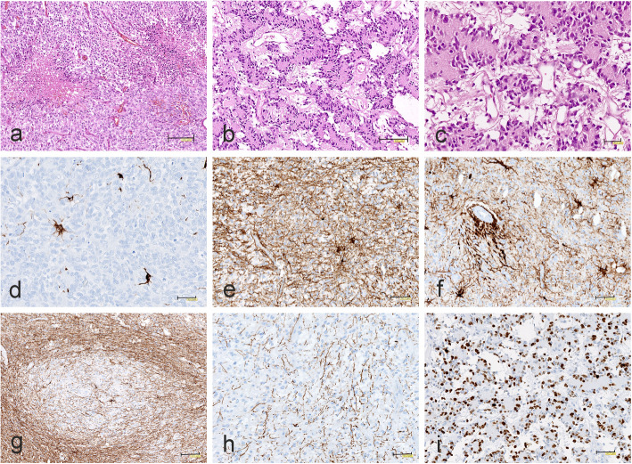
Morphology and immunophenotype of CNS-HGNT BCOR tumors. a Micronecrosis. b Papillary architecture and microcystic background. c Papillary structure with uniform, small cells arranged around eosinophilic acellular cores. d A few GFAP-immunopositive reactive astrocytes. e Strong GFAP immunoreactivity in reactive astroglial cells and tumor background. f Intense GFAP positivity around blood vessels. g Focal immunonegativity for GFAP. h A few tiny GFAP positive glial processes. i High Ki67 proliferation index in unusual malignant papillary neuroepithelial tumor. The scale bars: a - 150 μm; b, g - 100 μm; c - 30 μm; d-f, h, i - 50 μm

