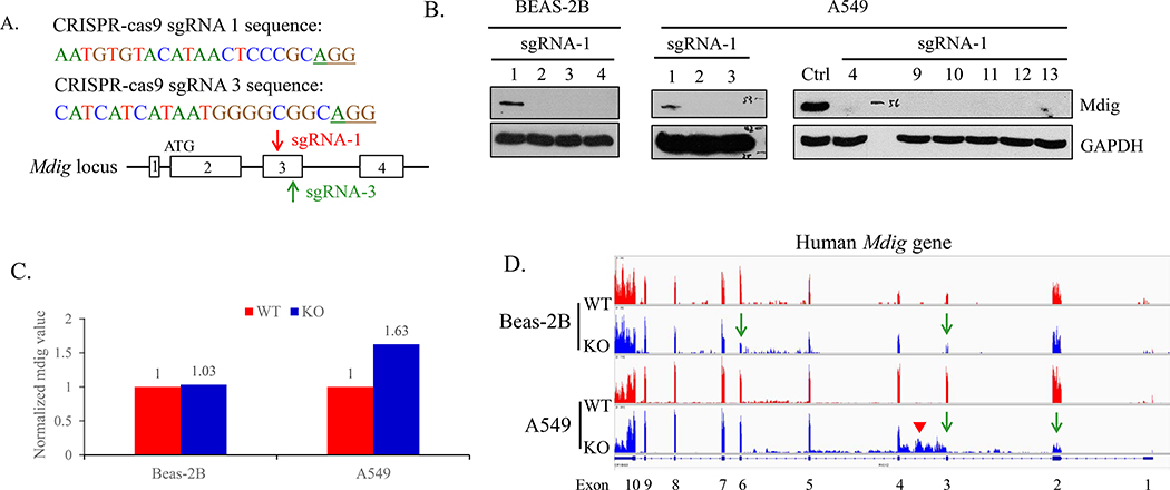
Fig 1.
Knockout of mdig by sgRNAs. A. Sequence and schematic of sgRNAs targeting the third exon of Mdig gene. The red arrow indicated the sgRNA-1, and the green arrow pointed sgRNA-3. B. Western blotting showing the mdig protein expression in BEAS-2B clones and A549 clones. C. Normalized mdig value from RNA sequencing (RNA-seq) of WT (red) and KO (blue) of BEAS-2B and A549 clones. D. RNA-seq showing the reads of mdig in WT (red) and KO (blue) of BEAS-2B and A549 clones.

