Fig. 2.
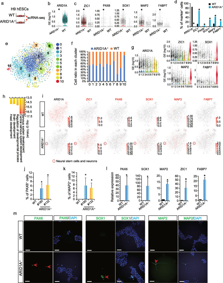
scRNA-seq reveals loss-of-ARID1A induces spontaneous neural differentiation in hESCs. a Undifferentiated WT and ARID1A−/− hESCs were collected for single-cell RNA sequencing (scRNA-seq). b ARID1A expression in WT and ARID1A−/− hESCs detected by scRNA-seq. *p < 2.4E−13 (Wilcoxon’s test). c Expression levels of neural stem cell markers (ZIC1, PAX6, SOX1) and neuron markers (MAP2, FABP7) in WT and ARID1A−/− hESCs. *p < 2.4E−13 (Wilcoxon’s test). d Percentage of cells expressing ARID1A, neural stem cell markers (ZIC1, PAX6, SOX1), and neuron markers (MAP2, FABP7) in WT and ARID1A−/− hESCs. *p < 1.1 E−14 (Fisher’s exact test). e Integrative analysis of scRNA-seq datasets from WT and ARID1A−/− hESCs. Cell clusters were visualized with t-distributed stochastic neighbor embedding (t-SNE). f Ratios of WT and ARID1A−/− hESCs in each cluster from integrative scRNA-seq data. g Violin plots of scRNA-seq data showing expression levels of ARID1A, neural stem cell markers (ZIC1, SOX1), and neuron markers (MAP2, FABP7) in each cluster. h GO biological process analysis for upregulated genes (ARID1A−/− vs. WT) in cluster 10 by Metacore software. i Feature plots of scRNA-seq data showing the distribution of cells positively expressing ARID1A, neural stem cell markers (ZIC1, PAX6, SOX1), and neuron markers (MAP2, FABP7) in all clusters. Red circle indicates the cluster 10 containing neural stem cells and neurons solely derived from in ARID1A−/− hESCs. j, k Flow cytometry data showing the percentage of PAX6+ (j) and MAP2+ (k) cells in WT and ARID1A−/− hESCs. All bars are shown as mean ± SD. n = 3, *p < 0.05 (KO vs. WT, an unpaired two-tailed t test with Welch’s correction). l Expression levels of neural-associated markers analyzed by qRT-PCR. All bars are shown as mean ± SD. n = 3, *p < 0.05 (KO vs. WT, an unpaired two-tailed t test with Welch’s correction). m Immunomicroscopy to detect neural stem cell markers (PAX6, SOX1) and neuron marker (MAP2) in WT and ARID1A−/− hESCs. Scale bar, 100 μm

