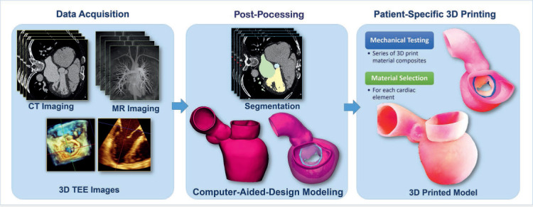
Figure 1.

3-dimensional (3D) printed model development workflow. Patient-specific 3D printed modeling starts with high-quality data acquisition from computed tomography (CT), magnetic resonance (MR) imaging, and 3D transesophageal echocardiography (TEE) images. The next step is imaging post-processing that includes geometry segmentation and computer-aided design modeling. The models are 3D printed using multiple materials that were previously selected based on mechanical testing.
