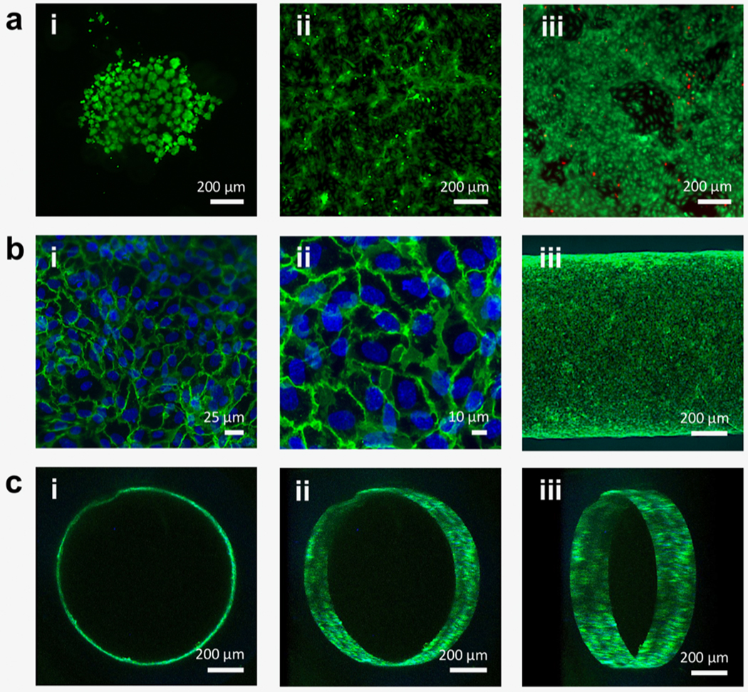
Figure 3.

Fluorescence and confocal micrographs of endothelial cell-seeded, cross-linked hydrogel constructs. (a; i) HUVECs seeded on cross-linked unmodified F127-BUM hydrogel (disc) after 24 h in culture. Cells stained with calcein AM (green) for visualization show aggregation and rounded morphology, indicating lack of adhesion. Percent coverage was determined to be 20% ± 9% (SD; n = 3). This image is representative of the three replicates. (a; ii) HUVECs seeded on cross-linked, collagen I-treated F127-BUM hydrogel with collagen I additive (disc) after 24 h in culture. Cells stained with calcein AM for visualization show spreading, indicating adhesion. Percent coverage was determined to be 52% ± 8% (SD; n = 3). This image is representative of the three replicates. (a; iii) Viability assay (calcein AM, green/ethidium homodimer-1, red) of HUVECs seeded on cross-linked, collagen I-treated F127-BUM hydrogel with collagen I additive (disc) after 72 h in culture. Green indicates live cells and red indicates dead cells; cell viability is high. This image is representative of three replicates. (b; i, ii) Confocal micrographs of HUVECs seeded on the luminal surfaces of tubes composed of cross-linked, collagen I-treated F127-BUM hydrogel with collagen I additive. Cells stained with DAPI (blue: nuclei) and labeled via indirect immunofluorescence (green: CD31, interendothelial junction marker) exhibit characteristic cobblestone morphology. These images are representative of six replicates. (b; iii) Identical treatment to (b; i, ii). Entire width of tube section is visualized, showing good cell coverage. This image is representative of six replicates. (c; i, ii, iii) Identical treatment to (b; i, ii, iii). Luminal surface of tube is visualized via confocal microscopy showing good cell coverage. These images are representative of six replicates.
