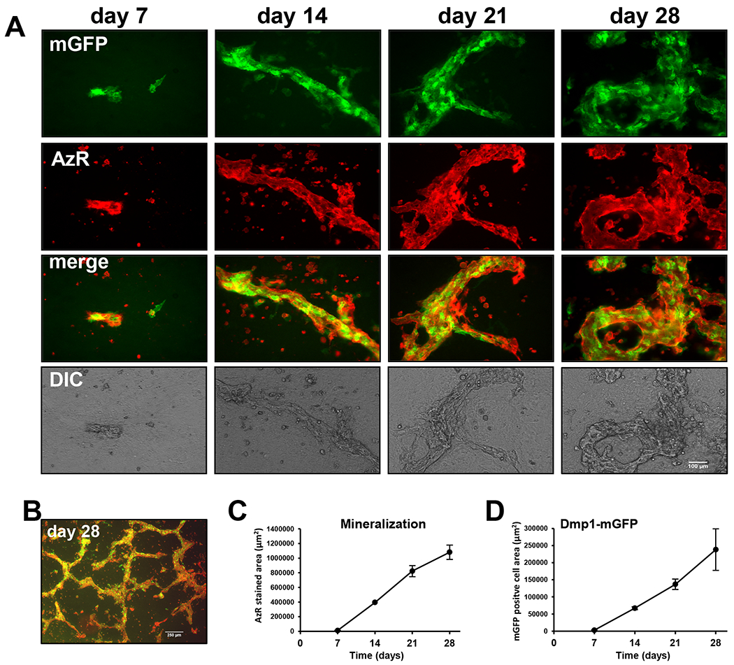
Figure 1: OmGFP66 cells form mineralized bone-like trabeculae over 7-28 days in culture.

A) Time course in OmGFP66 cells showing expression of the Dmp1-mGFP reporter (green) and alizarin red fluorescence (red) to monitor calcium deposition. Note the initial formation of small bone-like structures containing mineral and Dmp1-mGFP-positive cells by day 7. These bone-like structures increase in number and size over 28 days, bar = 100μm. B) Lower power view showing the trabecular-like nature of the bone mineral, bar = 250μm. C, D) Quantitation of alizarin red stained area (C) and Dmp1-mGFP positive cell area (D) over 28 days (data are mean ± SEM, n= 4).
