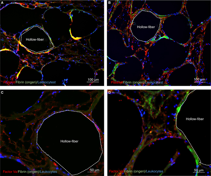
FIGURE 4.
Histochemistry of hollow-fiber oxygenators: Representative images from low (0.3 L/min) and high (0.7 L/min) oxygenators with orange dotted line outlying the inside of a representative gas fiber. A, Color micrograph of a cross section of a low-flow oxygenator with platelets (red), fibrinogen (green), and leukocytes (blue). B, Color micrograph of a cross section of a high-flow oxygenator with platelets (red), fibrinogen (green), and leukocytes (blue). C, Color micrograph of a cross section of a low-flow oxygenator with factor Va (red) and leukocytes (blue). D, Color micrograph of a cross section of a low-flow oxygenator with factor Va (red), fibrinogen (green), and leukocytes (blue)

