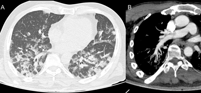
Figure 2.

Axial tomography images of the chest showing ground-glass opacities, some with thickening of interlobular septa, featuring mosaic paving, and some consolidations (A).
CTA of the chest, oblique reconstruction, demonstrating filling defects in arterial branches to the right lower lobe compatible with thromboembolism (B)
