Figure 4.

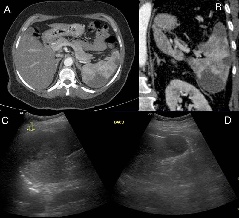
Axial and coronal abdominal CTA images showing wedge-shaped areas close to the convex face of the spleen compatible with splenic infarctions (A, B). Ultrasound images showing hypoechoic areas suggestive of splenic infarction (C, D)

Axial and coronal abdominal CTA images showing wedge-shaped areas close to the convex face of the spleen compatible with splenic infarctions (A, B). Ultrasound images showing hypoechoic areas suggestive of splenic infarction (C, D)