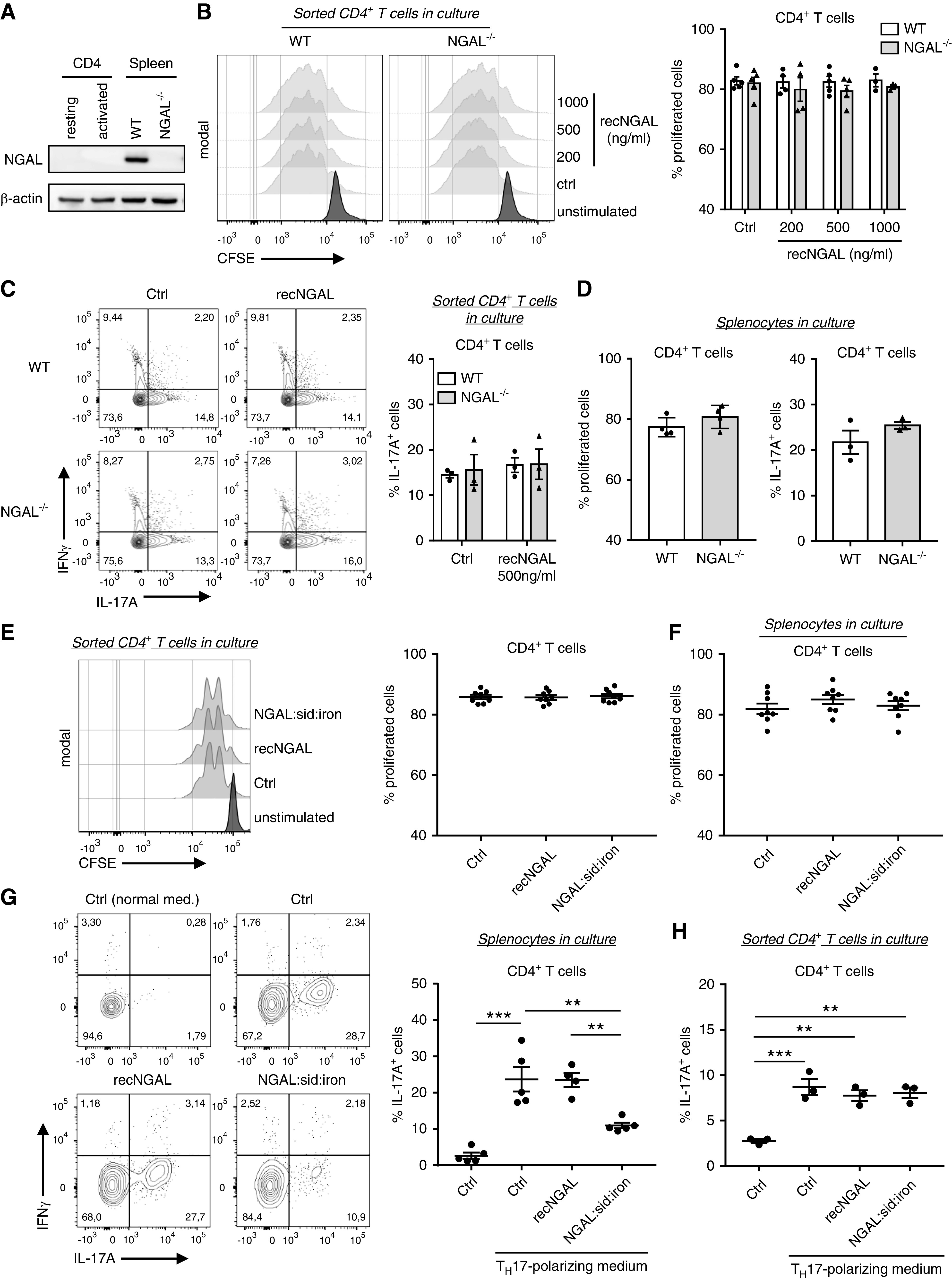
Figure 6.

Isolated WT and NGAL−/− CD4+ T cells show similar proliferation and TH17 polarization whereas siderophore iron–loaded NGAL inhibited TH17 polarization in splenocyte cultures. (A) Representative Western blot of NGAL and β-actin (loading control) expression in resting and CD3/CD28 Dynabeads-activated (activated) CD4+ cells isolated from WT mice spleens and in total splenocytes from WT and NGAL−/− mice. (B) CD4+ T cells isolated from spleens of WT and NGAL−/− mice were labeled with carboxyfluorescein succinimidyl ester (CFSE) and incubated with CD3/CD28 dynabeads in the absence or presence of the indicated recombinant murine NGAL concentrations. After 72 hours in culture, proliferation was assessed by flow cytometry. Representative histograms and the corresponding percentages of proliferated cells are shown. (C) Isolated CD4+ T cells from spleens of WT and NGAL−/− mice were incubated with CD3/CD28 dynabeads in TH17 polarization medium in absence or presence of 500 ng/ml recombinant murine NGAL for 6 days and analyzed by flow cytometry. Representative histograms and corresponding percentages of TH17 T cells (IL-17A+IFNγ −) are shown. (D) Proliferation and polarization assays were performed with total splenocytes from WT and NGAL−/− mice in culture. Proliferation and polarization of WT and NGAL−/− CD4+ T cells were found to be similar. (E) Proliferation of CD4+ T cells isolated from spleens of WT −/− mice was measured in the absence or presence of 1 μg/ml of either normal NGAL or iron siderophore–loaded recombinant murine NGAL. Representative histograms and the corresponding percentages of proliferated cells are shown. (F) Total splenocytes from WT mice were prepared and cultured as in (E). Proliferation of gated WT CD4+ T cells were similar in all conditions after 72 hours in culture. (G) Polarization assay of splenocytes from WT mice were performed as in (B) in absence or presence of 1 μg/ml of either normal NGAL or iron siderophore–loaded NGAL. Iron siderophore–loaded NGAL significantly reduced TH17 polarization compared with control conditions and normal NGAL, respectively. Representative histograms and corresponding percentages of TH17 cells (IL-17A+IFNγ−) are shown. (H) TH17 polarization assay under the same conditions as in (G) was performed with CD4+ T cells isolated from spleens of WT mice. Iron siderophore–loaded NGAL had no direct effect on CD4+ T cells during TH17 polarization. Percentages of TH17 T cells (IL-17A+IFNγ−) are shown. Ctrl, control; recNGAL, recombinant NGAL; sid, siderophore. **P<0.01; ***P<0.001.
