
Figure 5.
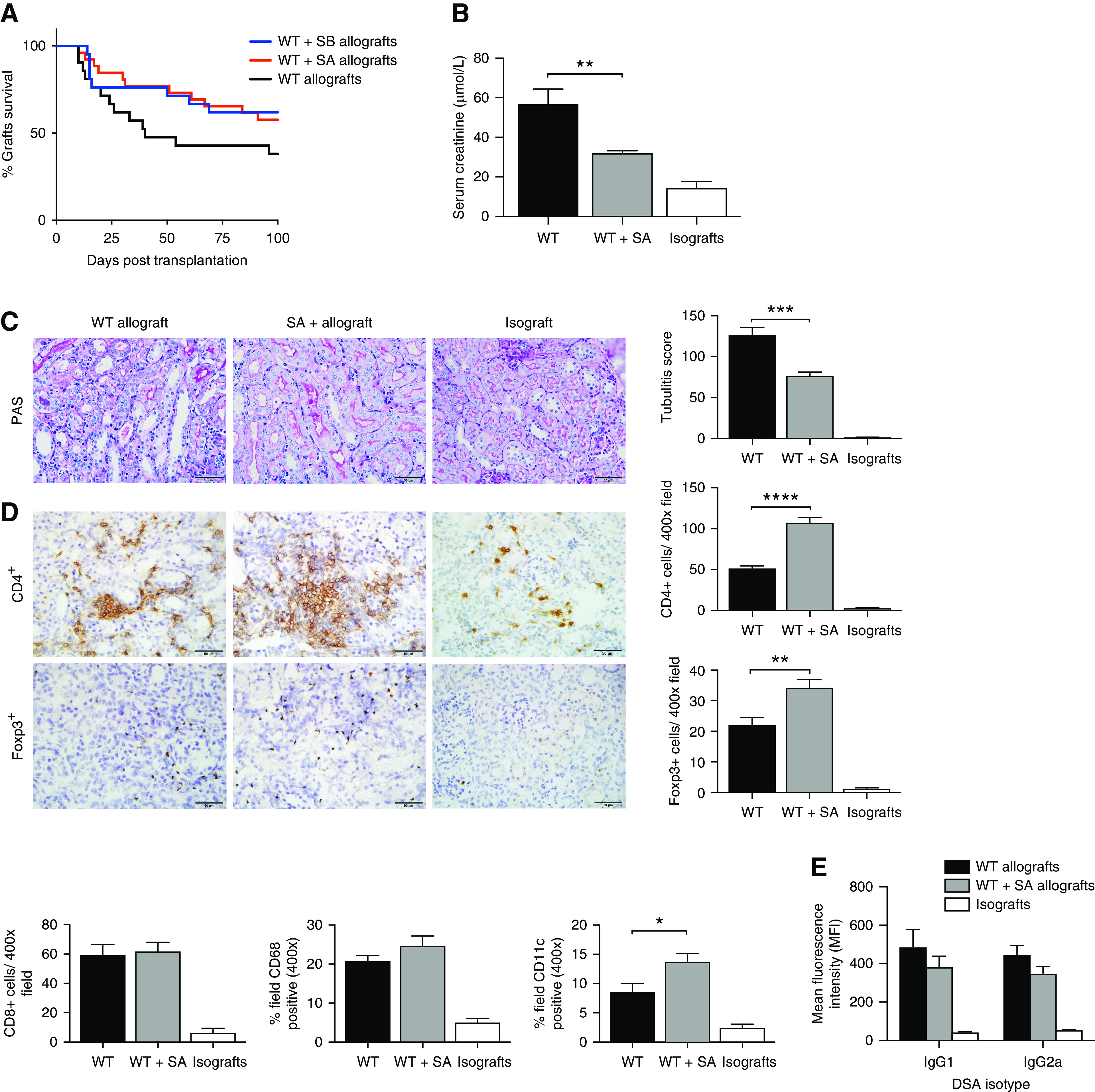
SCFA supplementation attenuated acute allograft rejection. (A) WT+SA allografts (n=27) and WT+SB allografts (n=21) exhibited a trend toward improved survival compared with WT allografts (n=21), without reaching significance. At day 14 post-transplant, WT+SA allografts (n=9) demonstrated improved graft function with lower serum creatinine (P<0.05) (B), and less tubulitis (C) compared with WT allografts (n=7) (P<0.01). (D) Representative photomicrographs and analysis of cellular infiltrate by IHC showed a marked increase in CD4+ and Foxp3+ T cells, as well as CD11c+ cells in WT+SA allografts compared with WT allografts (isografts n=5). (E) Serum donor-specific antibody titers were evaluated at 14 days post-transplantation. WT (n=13) and SA-supplemented (n=9) allograft mice demonstrated elevated donor-specific IgG1 and IgG2a compared with isografts (n=5). Scale bar, 50.0 μm. Data are shown as the mean±SEM. Statistical analysis by one-way ANOVA with Tukey post hoc analysis. *P<0.05; **P<0.01; ***P<0.001; ****P<0.0001.