
Figure 1.
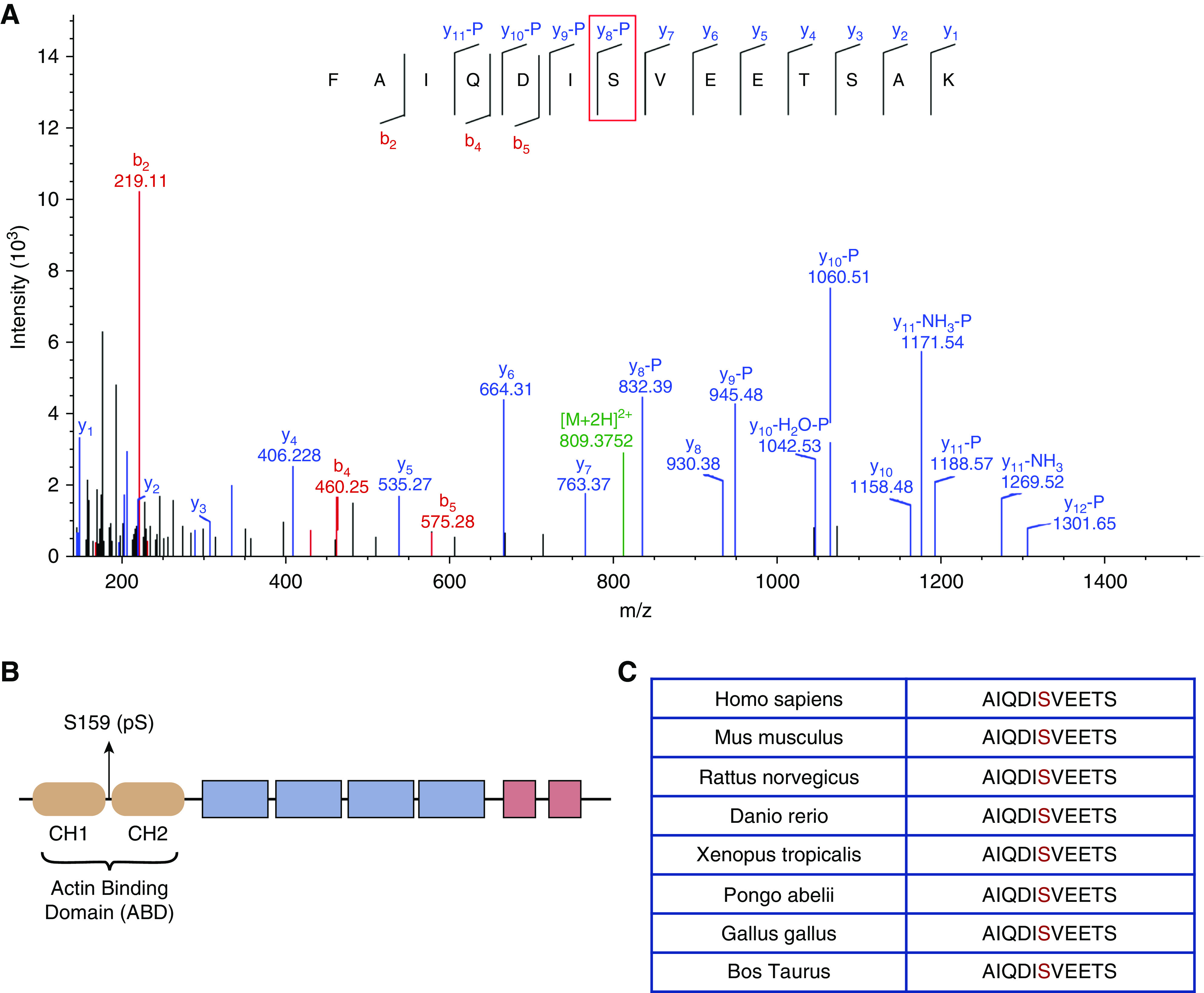
Phosphorylation of ACTN4 is detected at conserved. S159. (A) The representative tandem mass spectrometry (MS2)spectrum of the phosphopeptide (153FAIQDISVEETSAK166) showing S159 phosphorylation. The MS2 spectrum of the peptide at m/z=809.3752 (z=2) is shown in green in (A). Detection of specific y (blue) and b (red) fragment ions allowed identification of the peptide sequence 153FAIQDISVEETSAK166 and assignment of phosphorylation site to S159. Specifically, the presence of the y(8)-98 fragment ion confirms the site of phosphorylation at S159. (B) Functional domains of the human ACTN4 protein. The ABD consists of CH1 and CH2 domains. S159 is located in the linker region (amino acids 157–164) between CH1 and CH2 domain. (C) The S159 phosphorylation site (±5 amino acids from S159) is evolutionarily conserved across species.